The new technique will make the images clearer and you’re able to rotate 360 degrees. The researchers hope that this is not only allowing parents to visualize their future children, but it will also help researchers to better understand foetal anatomy.
The researchers were able to use the technique to visualize and make 3D models of 25 foetuses in their research. In two cases the technique didn’t work, because of the levels of amniotic fluid. These levels were too low for the researchers to get images of the foetus.
But in the cases where the technique worked, “we found these images more real, and the possibility that we can see in 360 degrees presents us with a greater interaction with the exam,” said study co-author Dr. Heron Werner Jr. from Clinical Diagnostic Imaging said. Werner and his colleagues recently presented the technique at the annual meeting of the Radiological Society of North America.
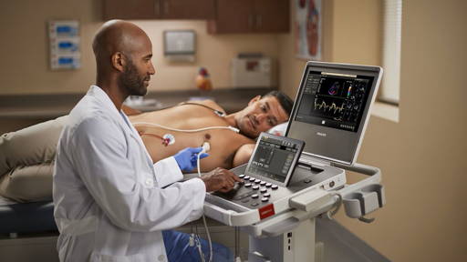
First 3D models of a foetus are produced via MRI and ultrasound techniques or in a combination of the two. This means women would undergo a similar imaging exam as the regular obstetric ultrasound, or MRI. But then, the researchers use frames of these images, in sequence, to make a 3D model of the foetus, according to the researchers.
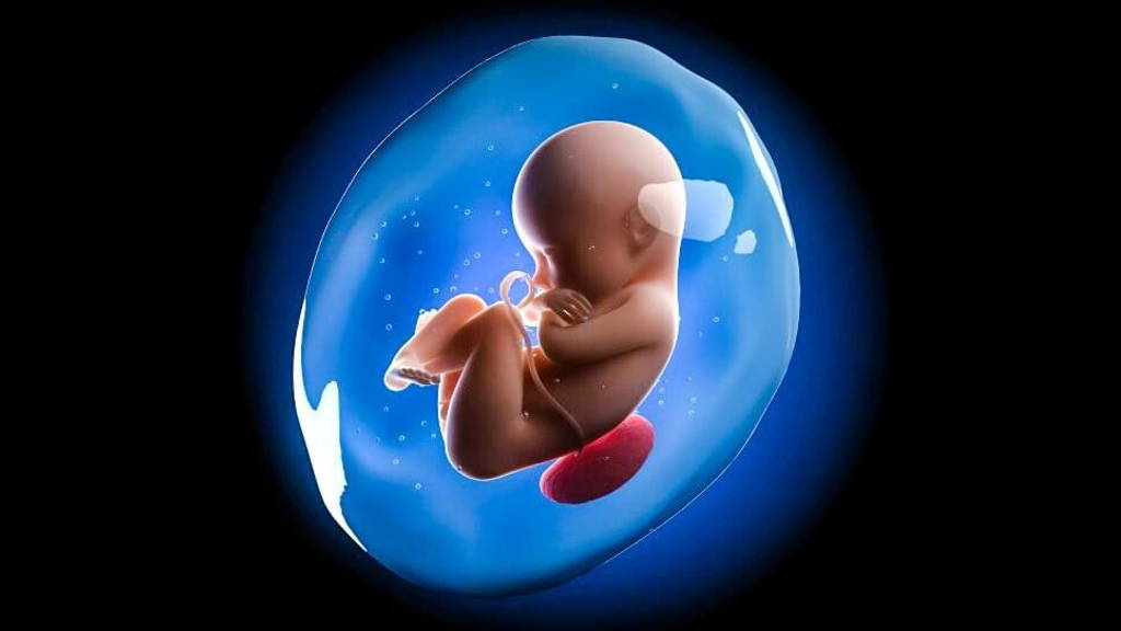
Then a physician can choose to focus on specific body parts of the foetus for the reconstruction. Finally, the parents-to-be can view the final image — which can include the inside of the womb, the umbilical cord and the placenta along with the fetus — through a virtual reality device.
The researchers used Oculus Rift 2 for their virtual reality headset. They found that it is possible to not only see their foetus all around, but they will also be able to hear the fetal heartbeat, by way of the ultrasound.
Besides the fact that it will be a whole new experience for the future-parents, this technology can also provide new options for evaluation of the baby’s health and development. The scan allows users to see all of the internal structures as well as external features of the foetus.
In this research, the scans did not reveal any health conditions in the foetuses that were not already known. But Werner explains that the technique improves the ability of multidisciplinary health care teams to work together and talk with family members about potential health problems. He also stated that the new technology can also help with planning pre- and postnatal surgeries.
The researchers were able to use the technique to visualize and make 3D models of 25 foetuses in their research. In two cases the technique didn’t work, because of the levels of amniotic fluid. These levels were too low for the researchers to get images of the foetus.
But in the cases where the technique worked, “we found these images more real, and the possibility that we can see in 360 degrees presents us with a greater interaction with the exam,” said study co-author Dr. Heron Werner Jr. from Clinical Diagnostic Imaging said. Werner and his colleagues recently presented the technique at the annual meeting of the Radiological Society of North America.
First 3D models of a foetus are produced via MRI and ultrasound techniques or in a combination of the two. This means women would undergo a similar imaging exam as the regular obstetric ultrasound, or MRI. But then, the researchers use frames of these images, in sequence, to make a 3D model of the foetus, according to the researchers.
Then a physician can choose to focus on specific body parts of the foetus for the reconstruction. Finally, the parents-to-be can view the final image — which can include the inside of the womb, the umbilical cord and the placenta along with the fetus — through a virtual reality device.
The researchers used Oculus Rift 2 for their virtual reality headset. They found that it is possible to not only see their foetus all around, but they will also be able to hear the fetal heartbeat, by way of the ultrasound.
Besides the fact that it will be a whole new experience for the future-parents, this technology can also provide new options for evaluation of the baby’s health and development. The scan allows users to see all of the internal structures as well as external features of the foetus.
In this research, the scans did not reveal any health conditions in the foetuses that were not already known. But Werner explains that the technique improves the ability of multidisciplinary health care teams to work together and talk with family members about potential health problems. He also stated that the new technology can also help with planning pre- and postnatal surgeries.