Graphene, atomically thin sheets of carbon, could be used for everything from nanoelectronics and aircraft de-icers, to batteries and bone implants. Recently, Engineers at the University of Wisconsin–Madison, published research on how to make and use researched graphene based microelectrocorticography (uECoG) arrays.
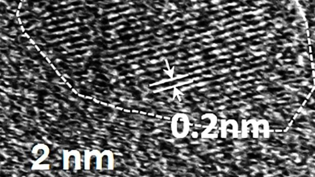
Another use for graphene may be as quantum dots sized contrast agents for magnetic resonance imaging (MRI), according to new research from Rice University. Quantum dots are microcopic, nano-scale objects that are small enough to penetrate cells and other biological items, that can be detected using various sensing technologies.
Rice University scientists have been working on overcoming this toxicity, using graphene-based quantum dots that consist of carbon, hydrogen, oxygen, and fluorine. Additionally, because different biomarkers can probably be attached to these quantum dots, one could produce contrast agents that show up only within certain types of tissues. If this pans out to be true, it may make spotting locations of tumors considerably easier than it is now.
“They have a lot of advantages compared with conventionally available contrast agents,” Rice researcher Sruthi Radhakrishnan said of the graphene-based quantum dots she has studied for the past two years. “Virtually all of the widely used contrast agents contain toxic metals, but our material has no metal. It’s just carbon, hydrogen, oxygen and fluorine, and in all of our tests so far it has shown no signs of toxicity.”
“There are tried-and-true methods for attaching biomarkers to carbon nanoparticles, so one could easily envision using these quantum dots to develop tissue-specific contrast agents,” Ajayan said. “For example, this method could be used to selectively target specific types of cancer or brain lesions caused by Alzheimer’s disease. That kind of specificity isn’t available with today’s contrast agents.”
The fluorinated graphene oxide quantum dots Radhakrishnan studies can be made in less than a day, but she spent two years perfecting the recipe for them. “It required a lot of optimization,” she said. “The recipe matters a lot.” Radhakrishnan said she plans to continue studying the material and hopes to eventually have a hand in proving that it is safe and effective for clinical MRI tests.
Another use for graphene may be as quantum dots sized contrast agents for magnetic resonance imaging (MRI), according to new research from Rice University. Quantum dots are microcopic, nano-scale objects that are small enough to penetrate cells and other biological items, that can be detected using various sensing technologies.
No toxic metals
MRI contrast agents shorten the amount of time it takes for tissues to realign and significantly improve the resolution of MRI scans. Almost all commercially available contrast agents are made from toxic metals like gadolinium, iron or manganese. This is a medical compromise between the benefits scans can provide and the negative effects of the substances that improve the quality of the scans.Rice University scientists have been working on overcoming this toxicity, using graphene-based quantum dots that consist of carbon, hydrogen, oxygen, and fluorine. Additionally, because different biomarkers can probably be attached to these quantum dots, one could produce contrast agents that show up only within certain types of tissues. If this pans out to be true, it may make spotting locations of tumors considerably easier than it is now.
“They have a lot of advantages compared with conventionally available contrast agents,” Rice researcher Sruthi Radhakrishnan said of the graphene-based quantum dots she has studied for the past two years. “Virtually all of the widely used contrast agents contain toxic metals, but our material has no metal. It’s just carbon, hydrogen, oxygen and fluorine, and in all of our tests so far it has shown no signs of toxicity.”
Targeting specific tissues
Pulickel Ajayan, the Rice materials scientist who is directing the work, said the fluorinated graphene oxide quantum dots could be particularly useful as MRI contrast agents because they could be targeted to specific kinds of tissues.“There are tried-and-true methods for attaching biomarkers to carbon nanoparticles, so one could easily envision using these quantum dots to develop tissue-specific contrast agents,” Ajayan said. “For example, this method could be used to selectively target specific types of cancer or brain lesions caused by Alzheimer’s disease. That kind of specificity isn’t available with today’s contrast agents.”
The fluorinated graphene oxide quantum dots Radhakrishnan studies can be made in less than a day, but she spent two years perfecting the recipe for them. “It required a lot of optimization,” she said. “The recipe matters a lot.” Radhakrishnan said she plans to continue studying the material and hopes to eventually have a hand in proving that it is safe and effective for clinical MRI tests.