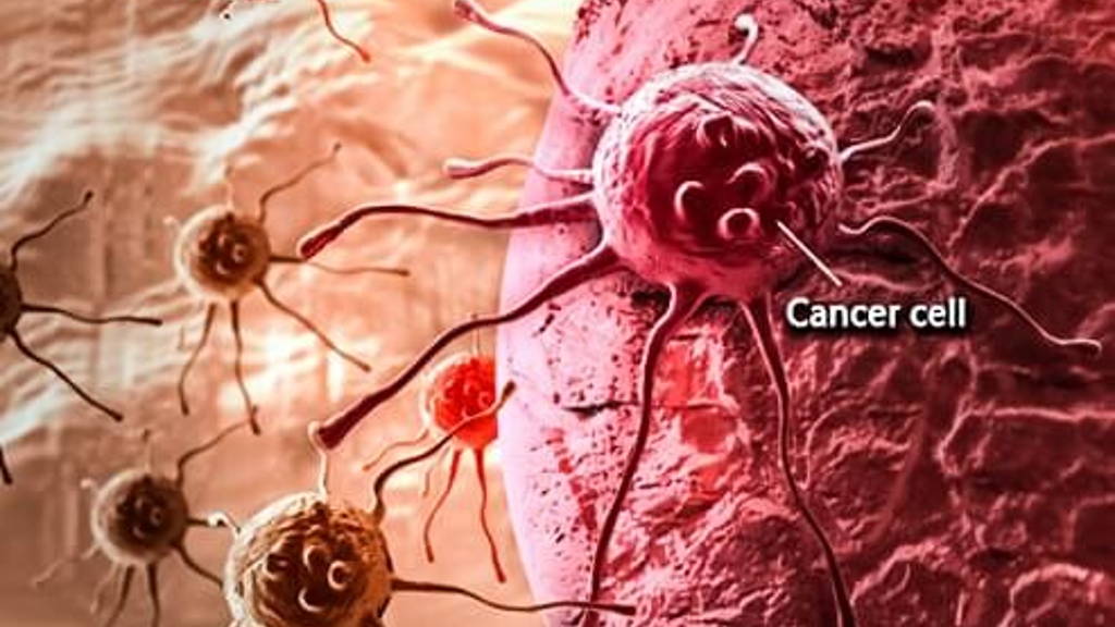
Researchers from Brown University have developed a new image analysis technique to distinguish two key cancer cell types associated with tumor progression. The approach could help in pre-clinical screening of cancer drugs and shed light on a cellular metamorphosis that is associated with more malignant and drug-resistant cancers.
The epithelial-mesenchymal transition, or EMT, is a process by which more docile epithelial cells transform into more aggressive mesenchymal cells. Tumors with higher numbers of mesenchymal cells are often more malignant and more resistant to drug therapies. The new technique combines microscopic imaging with a machine learning algorithm to better identify and distinguish between the two cell types in laboratory samples.
Susan Leggett, a doctoral student in Brown’s pathobiology graduate program and lead author of a paper describes the technique: “We know that there are these different cell types interacting within tumors and that therapeutics can target these cells differently. We’ve developed a model that can pick out these cell types automatically and in an unbiased way. We think this could help us better understand how these different cell types respond to drug treatment.”
In general, the epithelial and mesenchymal cell types can be distinguished by their shapes. Epithelial cells are more compact in appearance, while mesenchymal cells appear more elongated and spindly, both in their overall appearance and in the appearance of their nuclei.
In this training a epithelial cell line, cultured in a petri dish, served as a model for human breast cancer. The researchers activated a transcription factor called Snail -well known to cause these cells to quickly undergo an extreme form of EMT. Those cells, imaged before and after the transition, served as a training set to teach the algorithm to distinguish between the two cell types.
The researchers showed that, after training, the algorithm was able to categorize individual cells as either epithelial or mesenchymal with greater than 92 percent accuracy. Further experiments where used to refine this proces.
The team has already been studying whether Taxol, a chemo agent, promotes the epithelial–mesenchymal transition in cells it does not kill. The experiment found that while sub-lethal Taxol created a range of cell shapes, more than 70 percent of those could be classified by the algorithm as mesenchymal.
It’s a preliminary finding that will require much more study to fully understand, Wong states. But it could shed light on how tumors become resistant to Taxol and other drugs. With more development, the researchers think their technique could provide a new means to screen the effectiveness of cancer drugs.
About the study
Legget and Wong’s coauthors on the study were Jea Yun Sim, Jonathan Rubins, Zachary Neronha and Evelyn Kendall Williams, all from Brown. The research was supported by the National Institutes of Health (5T32ES007272-24), the COBRE Center for Cancer Research Development at Rhode Island Hospital (1P30GM110759-01A1), a Rhode Island Foundation Medical Research Grant, Jason and Donna McGraw Weiss and Brown University.
Read about the study in journal Integrative Biology: Morphological single cell profiling of the epithelial–mesenchymal transition (http://pubs.rsc.org/en/Content/ArticleLanding/2016/IB/C6IB00139D#!divAbstract)
The epithelial-mesenchymal transition, or EMT, is a process by which more docile epithelial cells transform into more aggressive mesenchymal cells. Tumors with higher numbers of mesenchymal cells are often more malignant and more resistant to drug therapies. The new technique combines microscopic imaging with a machine learning algorithm to better identify and distinguish between the two cell types in laboratory samples.
High speed vision system
The researchers’ high speed vision system outlines cells that just went through an epithelial–mesenchymal transition (EMT). Within tumors such cells lead to greater malignancy and increased drug resistance. The new imaging technique might allow researchers and clinicians to quickly identify if a particular drug is preventing the transition process or whether it is letting it go its course.Susan Leggett, a doctoral student in Brown’s pathobiology graduate program and lead author of a paper describes the technique: “We know that there are these different cell types interacting within tumors and that therapeutics can target these cells differently. We’ve developed a model that can pick out these cell types automatically and in an unbiased way. We think this could help us better understand how these different cell types respond to drug treatment.”
In general, the epithelial and mesenchymal cell types can be distinguished by their shapes. Epithelial cells are more compact in appearance, while mesenchymal cells appear more elongated and spindly, both in their overall appearance and in the appearance of their nuclei.
To subtle shape differences
“It’s not hard to distinguish the two in the most extreme instances,” said Ian Y. Wong, assistant professor of engineering at Brown and the senior author of the research. “But sometimes the shape differences are subtle and it can be hard for humans to recognize the difference, which makes categorizing the two a bit arbitrary. The innovation here is that we can train a computer to pick out those more subtle variations.”In this training a epithelial cell line, cultured in a petri dish, served as a model for human breast cancer. The researchers activated a transcription factor called Snail -well known to cause these cells to quickly undergo an extreme form of EMT. Those cells, imaged before and after the transition, served as a training set to teach the algorithm to distinguish between the two cell types.
The researchers showed that, after training, the algorithm was able to categorize individual cells as either epithelial or mesenchymal with greater than 92 percent accuracy. Further experiments where used to refine this proces.
The team has already been studying whether Taxol, a chemo agent, promotes the epithelial–mesenchymal transition in cells it does not kill. The experiment found that while sub-lethal Taxol created a range of cell shapes, more than 70 percent of those could be classified by the algorithm as mesenchymal.
It’s a preliminary finding that will require much more study to fully understand, Wong states. But it could shed light on how tumors become resistant to Taxol and other drugs. With more development, the researchers think their technique could provide a new means to screen the effectiveness of cancer drugs.
About the study
Legget and Wong’s coauthors on the study were Jea Yun Sim, Jonathan Rubins, Zachary Neronha and Evelyn Kendall Williams, all from Brown. The research was supported by the National Institutes of Health (5T32ES007272-24), the COBRE Center for Cancer Research Development at Rhode Island Hospital (1P30GM110759-01A1), a Rhode Island Foundation Medical Research Grant, Jason and Donna McGraw Weiss and Brown University.
Read about the study in journal Integrative Biology: Morphological single cell profiling of the epithelial–mesenchymal transition (http://pubs.rsc.org/en/Content/ArticleLanding/2016/IB/C6IB00139D#!divAbstract)