Physicians have long used visual judgment of medical images to determine the course of cancer treatment. Has a tumor shrunk during the course of treatment over several months, or have new tumors developed? To answer questions like these, physicians often perform CT and MRI scans. Tumors are usually evaluated only visually, new tumors are often over-looked.
“Our program package increases confidence during tumor measurement and follow-up,” explains Mark Schenk from the Fraunhofer Institute for Medical Image Computing MEVIS in Bremen, Germany. “The software can, for example, determine how the volume of a tumor changes over time and supports the detection of new tumors.” The package consists of modular processing components and can help medical technology manufacturers automate progress monitoring.
The Fraunhofer developed method is unique in its use of deep learning, the institute writes in a press release. This new type of machine learning is stated to reach far beyond existing approaches. The method is helpful for image segmentation, during which experts designate exact organ outlines.
The new deep learning approaches promise improved results and should save physicians valuable time. To demonstrate their self-learning methods, Fraunhofer scientists trained the software with CT liver images from 149 patients. Results showed that the more data the program analyzed, the better it could automatically identify liver contours.
Deep learning methods can help reliably discover metastases and thus improve treatment outcomes.
Researchers focus on a combination of classical approaches and machine learning: “We wish to harness existing expertise to implement deep learning as effectively and reliably as possible,” stresses Harz. Fraunhofer MEVIS builds upon years of experience in practical application: for example, the algorithms for highly precise lung image registration have been integrated into several commercial medical software applications.
“Our program package increases confidence during tumor measurement and follow-up,” explains Mark Schenk from the Fraunhofer Institute for Medical Image Computing MEVIS in Bremen, Germany. “The software can, for example, determine how the volume of a tumor changes over time and supports the detection of new tumors.” The package consists of modular processing components and can help medical technology manufacturers automate progress monitoring.
Deep learning
Machine learning methods comb through large data sets and identify common features and patterns. After training, the system can then analyze and evaluate new data sets of the same type. These methods are often employed for the automatic recognition of images, text, and speech.The Fraunhofer developed method is unique in its use of deep learning, the institute writes in a press release. This new type of machine learning is stated to reach far beyond existing approaches. The method is helpful for image segmentation, during which experts designate exact organ outlines.
Time-consuming errors
Existing computer segmentation programs seek clearly defined image features such as certain gray values. “However, this can often lead to errors,” according to Fraunhofer researcher Markus Harz. “The software assigns areas to the liver that do not belong to the organ.” These errors must be corrected by physicians, a process which can often be quite time-consuming.The new deep learning approaches promise improved results and should save physicians valuable time. To demonstrate their self-learning methods, Fraunhofer scientists trained the software with CT liver images from 149 patients. Results showed that the more data the program analyzed, the better it could automatically identify liver contours.
Image registration
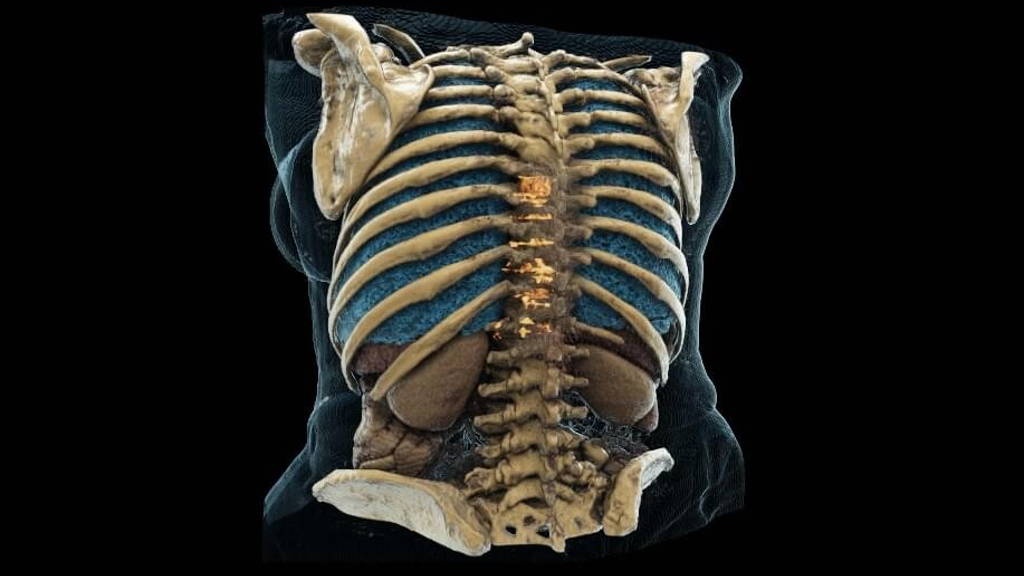
Another application of the approach is image registration, in which software aligns images from different patient visits so that physicians can easily compare them. Machine learning can aid the particularly difficult task of locating bone metastases in the torso in which hip bones, ribs, and spine are visible. Currently, these metastases are often overlooked due to time constraints in clinical practice.Deep learning methods can help reliably discover metastases and thus improve treatment outcomes.
Researchers focus on a combination of classical approaches and machine learning: “We wish to harness existing expertise to implement deep learning as effectively and reliably as possible,” stresses Harz. Fraunhofer MEVIS builds upon years of experience in practical application: for example, the algorithms for highly precise lung image registration have been integrated into several commercial medical software applications.