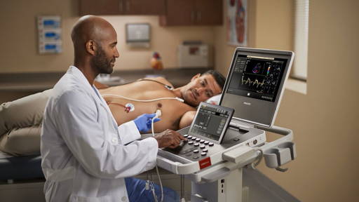
A team led by University of Utah electrical and computer engineering associate professor Rajesh Menon has developed a process called called ‘computational cannula microscopy’. It involves taking a needle about a quarter-millimeter in diameter and inserting it into the brain. Laser light shines through the needle and into the brain, illuminating certain cells like a flashlight.
The process has been developed for the benefit of medical researchers studying neurological disorders such as depression, obsessive-compulsive disorder and aggression. Menon and his team have been working with the U. of U.’s Nobel-winning researcher, Distinguished Professor of Biology and Human Genetics Mario Capecchi, and Jason Shepherd, assistant professor of neurobiology and anatomy.
The researchers genetically modify the mice so that only the cells they want to see glow under this laser light. The light from the glowing cells is captured by the needle and recorded by a standard camera. The captured light is run through a sophisticated algorithm developed by Menon and his team, which assembles the scattered light waves into a 2D or potentially, even a 3D picture.
Now that the process has been proven to work in animals, Menon believes it can potentially be developed for human patients, creating a simpler, less expensive and invasive method than endoscopes. “Although its much more complex from a regulatory standpoint, it can be done in humans, and not just in the brain, but for other organs as well, he says. “But our motivation for this project right now is to look inside the brain of the mouse and further develop the technique to understand fundamental neuroscience in the mouse brain.”
The research group has documented its process in a paper titled, “Deep-brain imaging via epifluorescence Computational Cannula Microscopy,” in the latest issue of Scientific Reports. The paper’s lead author is doctoral student Ganghun Kim. The paper’s co-authors include doctoral student Kyle Jenks and postdoctoral researchers Naveen Nagarajan and Elissa Pastuzyn.
The process has been developed for the benefit of medical researchers studying neurological disorders such as depression, obsessive-compulsive disorder and aggression. Menon and his team have been working with the U. of U.’s Nobel-winning researcher, Distinguished Professor of Biology and Human Genetics Mario Capecchi, and Jason Shepherd, assistant professor of neurobiology and anatomy.
The researchers genetically modify the mice so that only the cells they want to see glow under this laser light. The light from the glowing cells is captured by the needle and recorded by a standard camera. The captured light is run through a sophisticated algorithm developed by Menon and his team, which assembles the scattered light waves into a 2D or potentially, even a 3D picture.
Current methods
Typically, researchers must surgically take a sample of the animal’s brain to examine the cells under a microscope, or they use an endoscope that can be anywhere from 10 to 100 times thicker than a needle. “That’s very damaging,” Menon says of previous methods of examining the brain. “What we have done is to take a surgical needle that’s really tiny and easily put it into the brain as deep as we want and see very clear high-resolution images. This technique is particularly useful for looking deep inside the brain where other techniques fail.”Now that the process has been proven to work in animals, Menon believes it can potentially be developed for human patients, creating a simpler, less expensive and invasive method than endoscopes. “Although its much more complex from a regulatory standpoint, it can be done in humans, and not just in the brain, but for other organs as well, he says. “But our motivation for this project right now is to look inside the brain of the mouse and further develop the technique to understand fundamental neuroscience in the mouse brain.”
The research group has documented its process in a paper titled, “Deep-brain imaging via epifluorescence Computational Cannula Microscopy,” in the latest issue of Scientific Reports. The paper’s lead author is doctoral student Ganghun Kim. The paper’s co-authors include doctoral student Kyle Jenks and postdoctoral researchers Naveen Nagarajan and Elissa Pastuzyn.