The new ultrasound technology for neuroimaging - high-resolution, high-speed, safe and portable - was developed by physicist Prof. Tanter and his interdisciplinary team at the Ecole Supérieure de Physique et de Chimie Industrielles de la ville de Paris (ESPCI Paris) and at the Institut national de la santé et de la recherche médicale (INSERM U979 "Wave Physics for Medicine") in Paris, with the support of the European Research Council (ERC).
Tanter's technique is based on sonography, a method that relies on the generation of images by the bouncing of ultrasound waves. Commonly used by doctors - for instance during pregnancy - it had never been applied to neuroscience before as traditional ultrasound only allowed to image the blood flow in large blood vessels. To see the subtle neuronal activity of the smaller vessels of the brain, where the blood flow is less intense, an increased imaging sensitivity was indispensable. This was achieved by Tanter’s team by combining ultrafast imaging rates and dedicated processing algorithms.
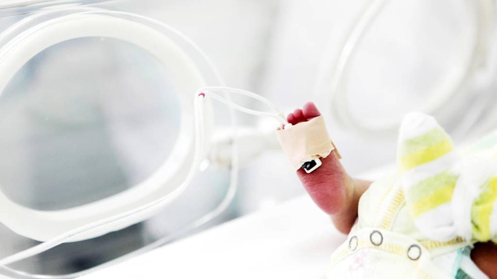
The recording of the cerebral activity of premature new-borns reported about is the result of a collaboration between the researcher and his colleague, Prof. Olivier Baud, paediatrician and responsible for the department of neonatology of Robert Debré paediatric hospital in Paris. It sets an unprecedented achievement, marking the entrance of ultrasound in the world of clinical neurosciences.
The machine was as big as a closet and needed 45 minutes to process all the data collected. Due to the technology state-of-the-art back of the 2000’s, the process was much slower than the one of the conventional Doppler method. Tanter had to wait until computer's had evolved sufficiently, in 2008, to build a ‘real-time’ machine.
The original development was nevertheless key, as it made possible to map the small and deep blood vessels of the brain with fast-speed, 50 times more sensitive, high resolution ultrasound. It also allowed to measure new and different parameters with direct clinical applications.
Tanter explains: "With this ultrafast imaging, we could see the mechanical vibrations of the body, like those produced by the heartbeat and the breathing. By focusing the fast-speed ultrasound beam on a specific organ, we were also able to create a vibration and "palpate", in a non-invasive way, a specific part inside the body, like a doctor would do with his hands if he could reach internal organs.” This "seismology" of the human body is key for diagnosis, Tanter says. “We can now, for instance, measure the stiffness of a lesion, identify its benign or malignant nature, and spot cancerous tissues.”
"With this technique we have observed the brains of preterm babies through the fontanel and can identify the underlying mechanisms for some pathological conditions or neurological disorders, such as neonatal seizures and haemorrhages", points out the EU-funded researcher grantee. Functional ultrasound imaging can likewise be used in adults, both in neurosurgery and for transcranial imaging, as new adaptive focusing techniques can overcome the strong aberrations induced by the skull bone on ultrasonic wave fronts.
In the near future, the resolution of fUS imaging could be further boosted thanks to a super-resolution ultrasound recently developed by the same research team. The whole functional activity of the brain could then be imaged at microscopic scales: a new world for neuroscience.
Non-invasive technique
The researchers say the technique is non-invasive and can be used in bedside conditions, in stark contrast with current tools such as the Functional Magnetic Resonance Imaging (fMRI), the Positron Emission Tomography (PET) and the Computed Tomography (CT). Despite their high level of performance, the latter can be big, noisy, time-consuming, expensive and very disrupting for patients.Tanter's technique is based on sonography, a method that relies on the generation of images by the bouncing of ultrasound waves. Commonly used by doctors - for instance during pregnancy - it had never been applied to neuroscience before as traditional ultrasound only allowed to image the blood flow in large blood vessels. To see the subtle neuronal activity of the smaller vessels of the brain, where the blood flow is less intense, an increased imaging sensitivity was indispensable. This was achieved by Tanter’s team by combining ultrafast imaging rates and dedicated processing algorithms.
The recording of the cerebral activity of premature new-borns reported about is the result of a collaboration between the researcher and his colleague, Prof. Olivier Baud, paediatrician and responsible for the department of neonatology of Robert Debré paediatric hospital in Paris. It sets an unprecedented achievement, marking the entrance of ultrasound in the world of clinical neurosciences.
New form of ultrasound imaging
Tanter's first attempt to boost ultrasound technology was back in 1999. With his team, he pioneered ultrafast ultrasound imaging based on plane wave transmission, instead of the usual sequential focused beams transmissions.The machine was as big as a closet and needed 45 minutes to process all the data collected. Due to the technology state-of-the-art back of the 2000’s, the process was much slower than the one of the conventional Doppler method. Tanter had to wait until computer's had evolved sufficiently, in 2008, to build a ‘real-time’ machine.
The original development was nevertheless key, as it made possible to map the small and deep blood vessels of the brain with fast-speed, 50 times more sensitive, high resolution ultrasound. It also allowed to measure new and different parameters with direct clinical applications.
Tanter explains: "With this ultrafast imaging, we could see the mechanical vibrations of the body, like those produced by the heartbeat and the breathing. By focusing the fast-speed ultrasound beam on a specific organ, we were also able to create a vibration and "palpate", in a non-invasive way, a specific part inside the body, like a doctor would do with his hands if he could reach internal organs.” This "seismology" of the human body is key for diagnosis, Tanter says. “We can now, for instance, measure the stiffness of a lesion, identify its benign or malignant nature, and spot cancerous tissues.”
Step forward with fUS technique
Tanter has now taken his research a step forward by developing a Functional Ultrasound (fUS) technique with massive computational power for the processing of fast ultrasound images, which can reveal the functional connectivity of the brain. This includes interactions and full sequences never observed before, such as epilepsy seizure propagation. Contrary to the fMRI, the functional ultrasound is light, portable, cheap, can be used in mobile animals and during surgery, thus offering new clinical applications and new paths for fundamental research."With this technique we have observed the brains of preterm babies through the fontanel and can identify the underlying mechanisms for some pathological conditions or neurological disorders, such as neonatal seizures and haemorrhages", points out the EU-funded researcher grantee. Functional ultrasound imaging can likewise be used in adults, both in neurosurgery and for transcranial imaging, as new adaptive focusing techniques can overcome the strong aberrations induced by the skull bone on ultrasonic wave fronts.
In the near future, the resolution of fUS imaging could be further boosted thanks to a super-resolution ultrasound recently developed by the same research team. The whole functional activity of the brain could then be imaged at microscopic scales: a new world for neuroscience.