A kidney usually roughly has the shape of a kidney bean – or the other way around – hence the name. In the case of Wes Nance, his kidneys had a different shape, plus they were positioned atypically inside his body. To make matters more difficult, he had roughly a dozen painful kidney stones. Removing these kidney stones would be complicated.
To find a solution, Jay Bishoff, MD, medical director of the Intermountain Medical Center Urological Institute in Salt Lake City, collaborated with a team from Intermountain Healthcare’s Innovation Lab. “In working with Dr. Bishoff, we were able to produce multiple advanced 3D images and models that not only helped him plan the best approach for Wes’ surgery, but he was able to use those images in the operating room to find and remove the kidney stones, despite Wes’s complex anatomy,” explains Billy Prows, manager of Intermountain Healthcare’s Innovation Lab.
• A 3D printed model of Wes’ kidneys and the placement of each kidney stone allowed them to examine his anatomy from multiple angles. The clear kidney wall, yellow urinary ducts, and blue kidney stones showed up clearly in the customized model.
• Using a laptop, Wes was shown a colorful 3D rendering of his kidneys in relation to his spine, pelvis, and other organs. The rendering could be digitally rotated and the organs could be removed from the screen to isolate the kidneys and show the location of his kidney stones.
• The team walked Wes through a 3D rendering of his anatomy on an iPad, which showed the layers Dr. Bishoff would have to go through to get to the kidney stones – skin, intestines, and the ducts in the kidneys.
“Imagine looking deep in a 40-inch hologram of a patient created from their standard MRI or CT images,” said Prows. “Without special glasses or a headset, it’s possible to use hand gestures to rotate and peel through the patient’s anatomy and see images that have the brightness and opacity of a standard flat-panel display.”
The team is now exploring other uses of the technologies to see if there are ways to create 3D images that can allow surgeons to practice surgery long before they actually step into the operating room.
To find a solution, Jay Bishoff, MD, medical director of the Intermountain Medical Center Urological Institute in Salt Lake City, collaborated with a team from Intermountain Healthcare’s Innovation Lab. “In working with Dr. Bishoff, we were able to produce multiple advanced 3D images and models that not only helped him plan the best approach for Wes’ surgery, but he was able to use those images in the operating room to find and remove the kidney stones, despite Wes’s complex anatomy,” explains Billy Prows, manager of Intermountain Healthcare’s Innovation Lab.
3d reconstructions
Wes Nance and his wife were shown some of the 3D reconstructions just prior to the actual surgery taking place. Among them:• A 3D printed model of Wes’ kidneys and the placement of each kidney stone allowed them to examine his anatomy from multiple angles. The clear kidney wall, yellow urinary ducts, and blue kidney stones showed up clearly in the customized model.
• Using a laptop, Wes was shown a colorful 3D rendering of his kidneys in relation to his spine, pelvis, and other organs. The rendering could be digitally rotated and the organs could be removed from the screen to isolate the kidneys and show the location of his kidney stones.
• The team walked Wes through a 3D rendering of his anatomy on an iPad, which showed the layers Dr. Bishoff would have to go through to get to the kidney stones – skin, intestines, and the ducts in the kidneys.
See-through anatomy
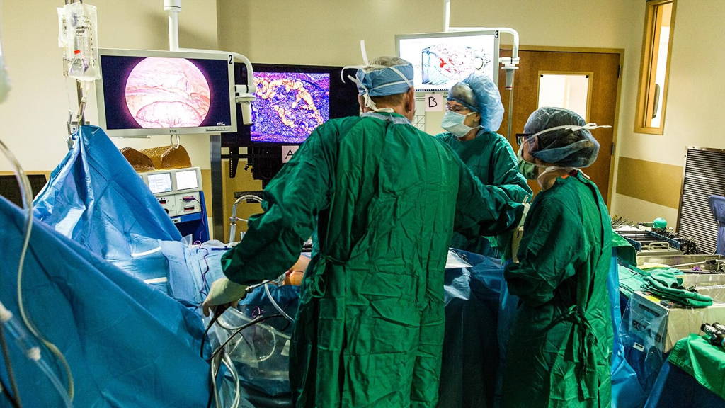
In the operating room, the third 3D reconstructed image of Nance's abdomen was paired with a 40-inch autosteroscopic display - glasses-free 3D television - which allowed Dr. Bishoff to see Nance's anatomy, layer by layer, throughout the surgery in a truly 3D view. This is, the IMC believes, the first time this type of glasses-free 3D technology has been used in a surgery.“Imagine looking deep in a 40-inch hologram of a patient created from their standard MRI or CT images,” said Prows. “Without special glasses or a headset, it’s possible to use hand gestures to rotate and peel through the patient’s anatomy and see images that have the brightness and opacity of a standard flat-panel display.”
Cutting-edge technology
“We were able to use these cutting-edge technologies to find and remove stones we otherwise wouldn’t have been able to find,” said Dr. Bishoff. “This allowed us to provide better care in a procedure that was simpler and less invasive than it would have been without these revolutionary new innovations.”The team is now exploring other uses of the technologies to see if there are ways to create 3D images that can allow surgeons to practice surgery long before they actually step into the operating room.